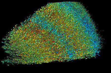
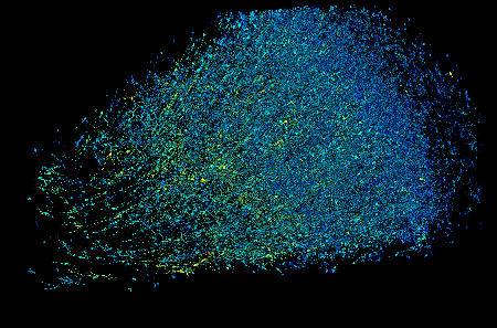
In the work published today, our team reports a new milestone: a synaptic-resolution reconstruction of a 1 cubic mm piece of human brain tissue. Our collaborators generated the dataset using a sample of brain tissue from the left anterior temporal lobe that was removed during brain surgery on a person with epilepsy. Lichtman’s team used a multibeam scanning electron microscope to gather high resolution images of more than 5,000 slices of tissue, each roughly 30 nanometers thick. Image acquisition alone took 326 days. Then our team’s tools stitched and aligned the image data, reconstructed the three dimensional structure of each cell, including its axons and dendrites, identified synaptic connections, and classified cell types. The reconstruction revealed several surprises.
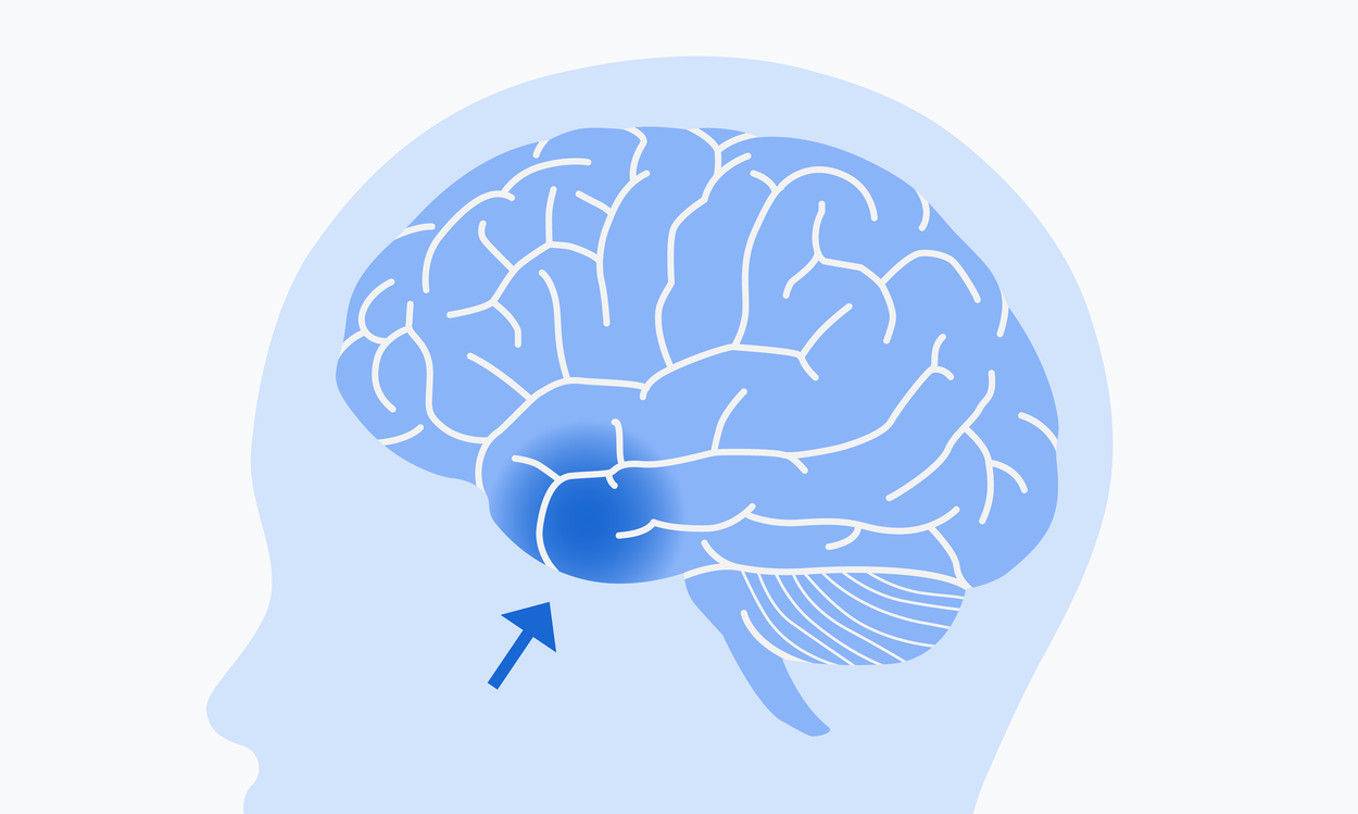 The brain tissue sample used in this research came from the left anterior temporal lobe.
For example, we found a class of rare but extremely powerful synaptic connections in which a pair of neurons may be connected by more than 50 individual synapses. While 96.5% of contacts between axons and their target cells have just one synapse, 0.092% have four or more synaptic connections. The conformation of these connections combined with refined statistical analysis that is part of the Science publication suggests that these powerful connections are not the result of chance, but rather that these pairs had a reason to be more strongly connected than is typical. Further study of these connections could reveal their functional role in the brain. Perhaps, for example, these strong connections are how the brain achieves particularly fast neural responses or how it encodes very important memories. […]
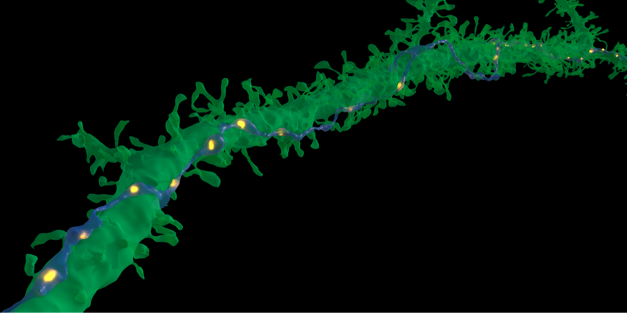 In very rare cases, a single axon (blue) made repeated synaptic connections (yellow) with a target neuron (green). The purpose of these very tight connections is not yet known. Credit: Google Research & Lichtman Lab (Harvard University). Renderings by D. Berger (Harvard University).